3D Visualization.
Using 3D models to create accurate main character of the spread/poster/infographics.
3D modeling and sculpting is the essential skill for biomedical communicators especially medical animators. As one of them, I model anatomical models and other biomedical forms based on scientific dataset and imaging.

Cellular and molecular visualization | Concept art | Game Design
Understand Alzheimer’s Disease
I am tasked to use Unreal Engine 5 to make a real-time game that is related to biomedical topics. Through playing this game, player will understand the basic concepts of Alzheimer’s disease, including the causes and consequences.
Player will use the gun to destroy the beta-amyloid plaques before they become bigger and attack the neurons. Their task is to stop the progression of Alzheimer’s disease!
Use of media: Unreal Engine 5, Zbrush, Adobe Substance Painter
Shoot down the beta-amyloid plaques before they invade your neurons!
Interactive Game Design


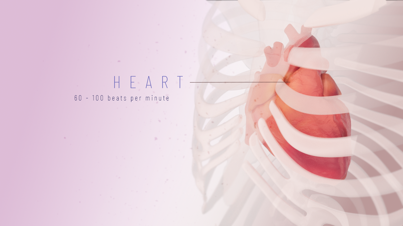
HUMAN HEART
Human heart pumps at about 60-100 beats per minute (bpm). Each pump consists of diastole and systole, where the ventricles and atria take turns to contract and relax, to create great pressure to push out the blood to the rest of the body.
3D modeling | Digital sculpting | Animation
To create an animation about the heart, a heart model is needed. To accurately depict the movement of the pumping of a heart, there was intensive research and trial and error to make it as realistic as it could be to reflect the real movement of one of the most important organ of our body.
Use of media: Cinema 4D, Autodesk Maya, Zbrush, Adobe Substance Painter, Redshift, After Effects
To model the heart, I had to go through a variety of 3D tools. It is complicated to model an organic and highly specific model. As the heart beats automatically and repeatedly as a steady motion, loop animation is created to allow the viewers to see the continuous pumping action.


Topology (Low-poly, high-poly)

Texture maps
Cellular and molecular visualization | Concept art
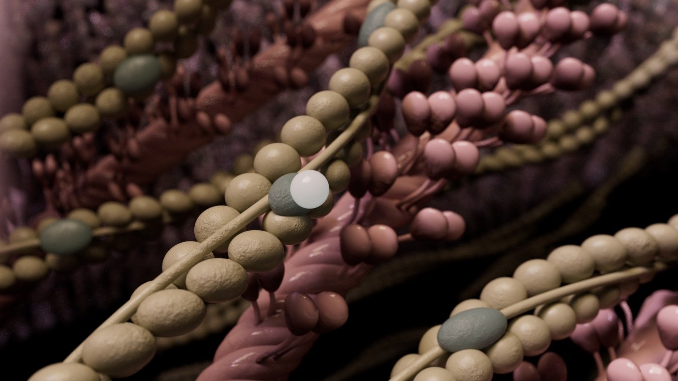
Myocardial microenvironment
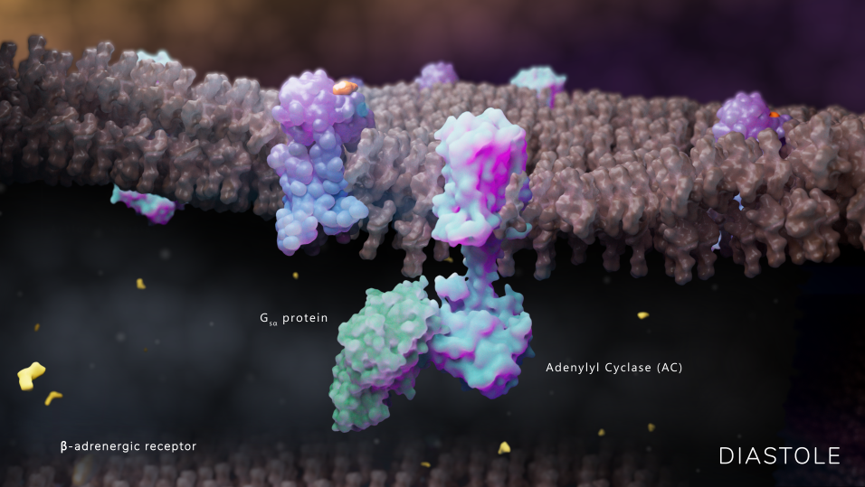
Explaining the process of excitation-contraction (EC) coupling
Myocardial microenvironment is referred to the environment inside the heart in a cellular and molecular level, something the human eye cannot see. To visualize different environmental scenes such as cardiomyocyte (cardiac muscle cell), movements across the cell membrane and actin and myosin actions in the cell, audience will be able to see the invisible,
This concept art project is dedicated to the creation of part of my thesis. The first part is to create and explain the normal excitation-contraction coupling (EC coupling) of the heart. To be simple, it means that why the heart can contract and relax, what do these actions lead to cardiac muscle contraction, and how do they turn back to normal. Also, questions like how are the electrical signals are being transduced and how the heart pumps the blood will be addressed.
Use of media: Autodesk Maya, Mudbox, Maxon Cinema 4D, Redshift, Red Giant, Adobe Photoshop, Illustrator, After Effects, Zbrush, Substance Painter, Protein data bank, ChimeraX.


Concept

Having a year of hand-on experience in 3D modeling and animation, I am comfortable to explore new 3D tools and the existing tools to create an animation series for my master’s thesis.
The heart is always fascinating. Its autonomy, its structure, its electrical pathways. I collaborate with cardiologist Dr. Bruce Lerman from Weill Cornell Medicine to create an animation series explaining the mechanism of cardiac excitation-contraction coupling (EC coupling), and the mechanism of disease of ventricular tachycardia triggered by delayed after-depolarizations (DADs) and early after-depolarizations (EADs). A lot of pathological mechanisms of cardiac diseases are visualized, but the molecular mechanisms and current flows of some of the diseases are still unknown and still waiting to be visualized, this is one of them.
This case study is completed for the first part of the series, which the target audience is the public who has little knowledge, and students who are studying biology/physiology. This 3-minute long animation provides an idea for audience how the heart pumps in a organ level, cellular level, and molecular level. I hope to link them together so audience would better understand how the biological hierarchy works.
The overall storytelling and storyline were developed and revised multiple times before storyboard development. I seek comments from content advisor and faculty professors for the storytelling approach and logical flow of the overall story.
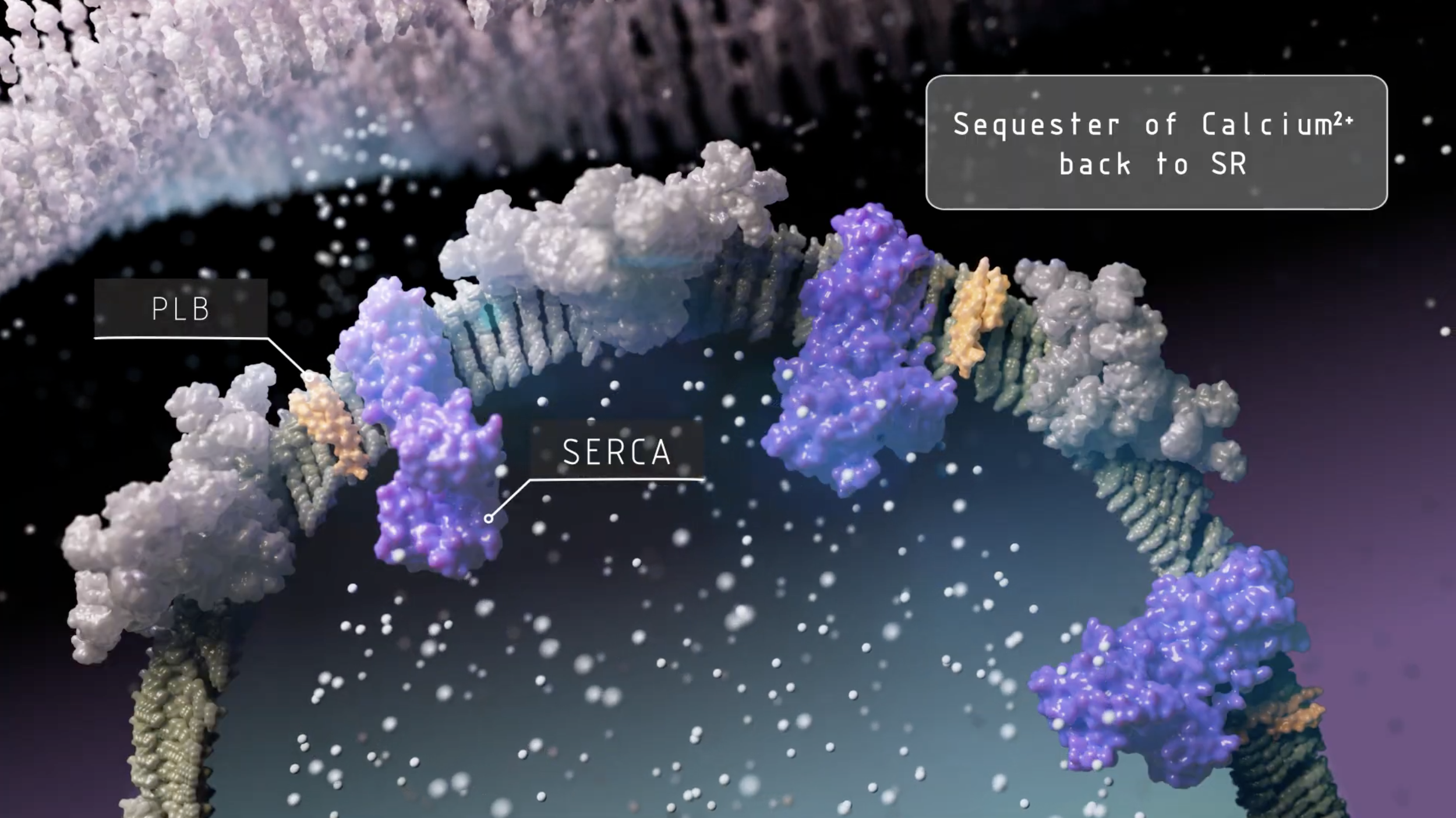
Crystal Structure of Endoplasmic Reticulum Ca2+-ATPase (SERCA)
Visual references


Didactic illustrations, histological slides, diagrams, and fluorescence-labelled images are references to produce highly accurate cardiomyocyte (cardiac muscle cell).
References from all range of media are used to develop the models and look development. As the topic is scientific and mysterious, a dark background could help the audience to focus on the saturated main character with high contrast in color. Data infographics are also referenced

Scientific references

Cryo-EM structure of RyR2
The structure of phospholamban bound to the calcium pump SERCA1a

Each molecular structure has their own unique and highly specific bondings and structure. Using protein visualization tools to extract the biological models are essential in molecular visualization to create accurate model and animation. Models are imported into Maya and Cinema 4D to be retouched, textured and later animated. Here are some examples:
Use of media: PDB, ChimeraX

Greyscale storyboards are developed for all important frames. Storytelling approach, camera position, transitioning all taken into consideration. Timecode, narration, captions are matched to each storyboard frame to provide the initial overall idea for the animation.
Storyboard


3D modeling | sculpting | Animation
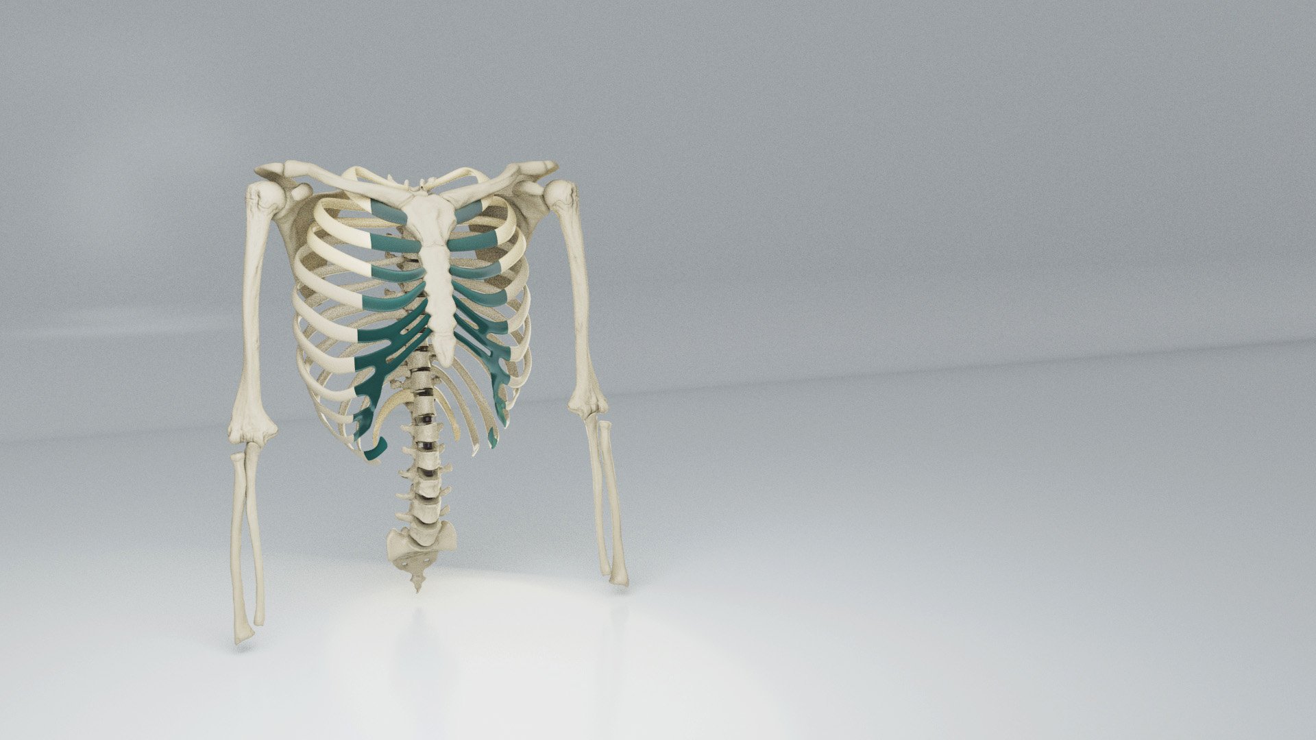
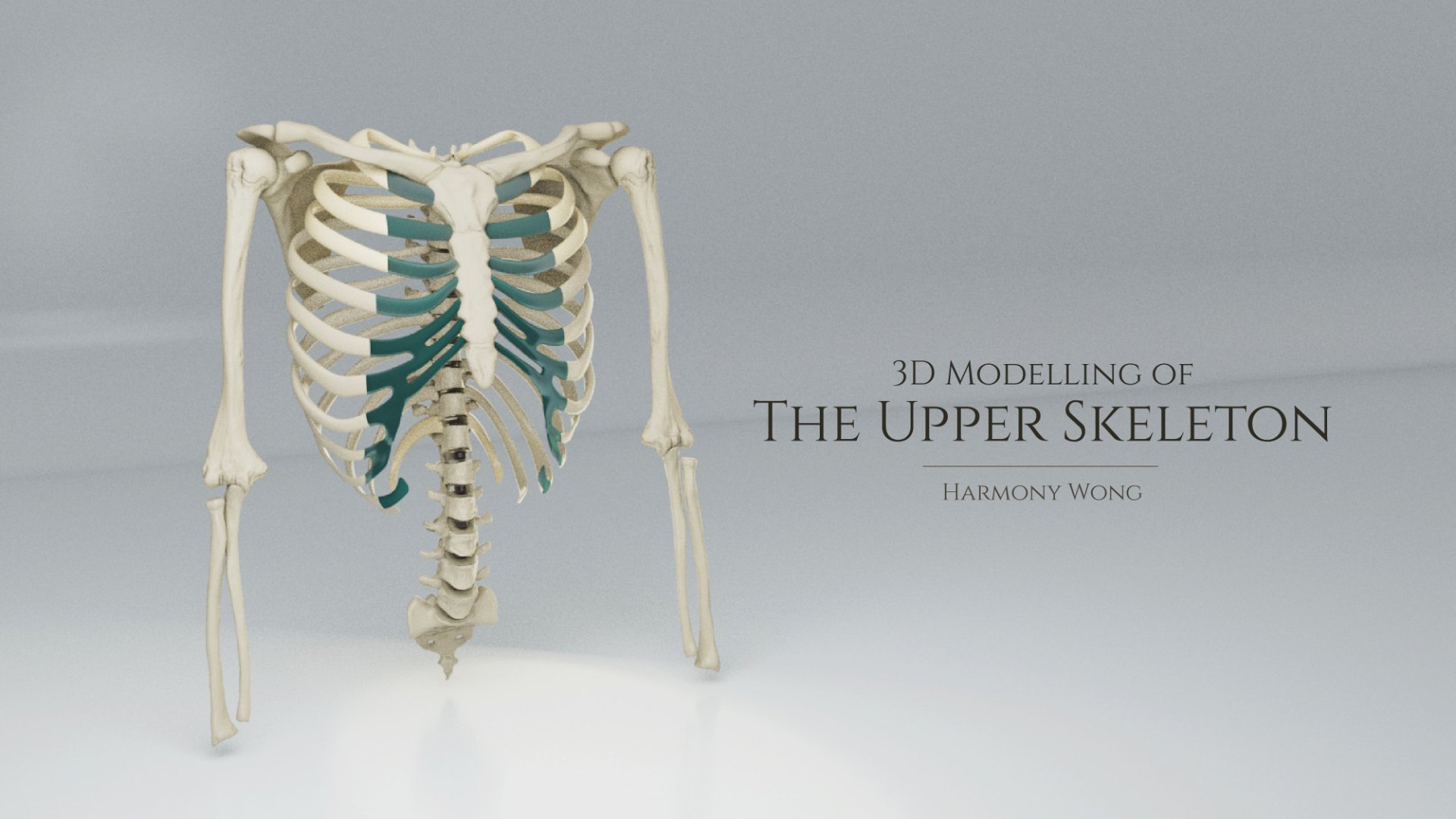
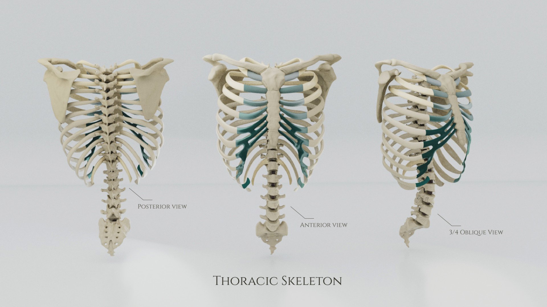
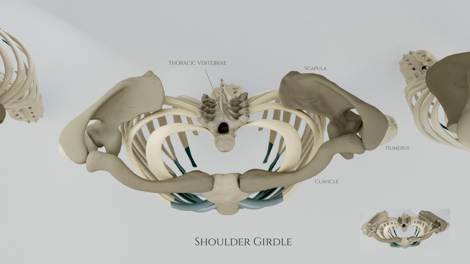
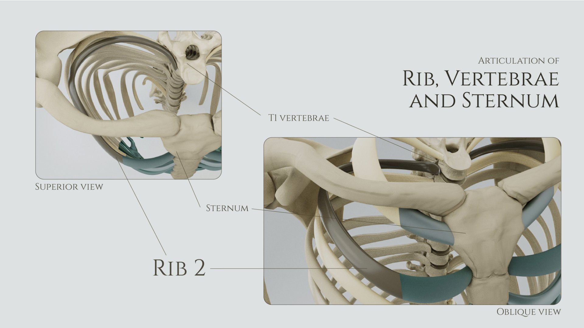
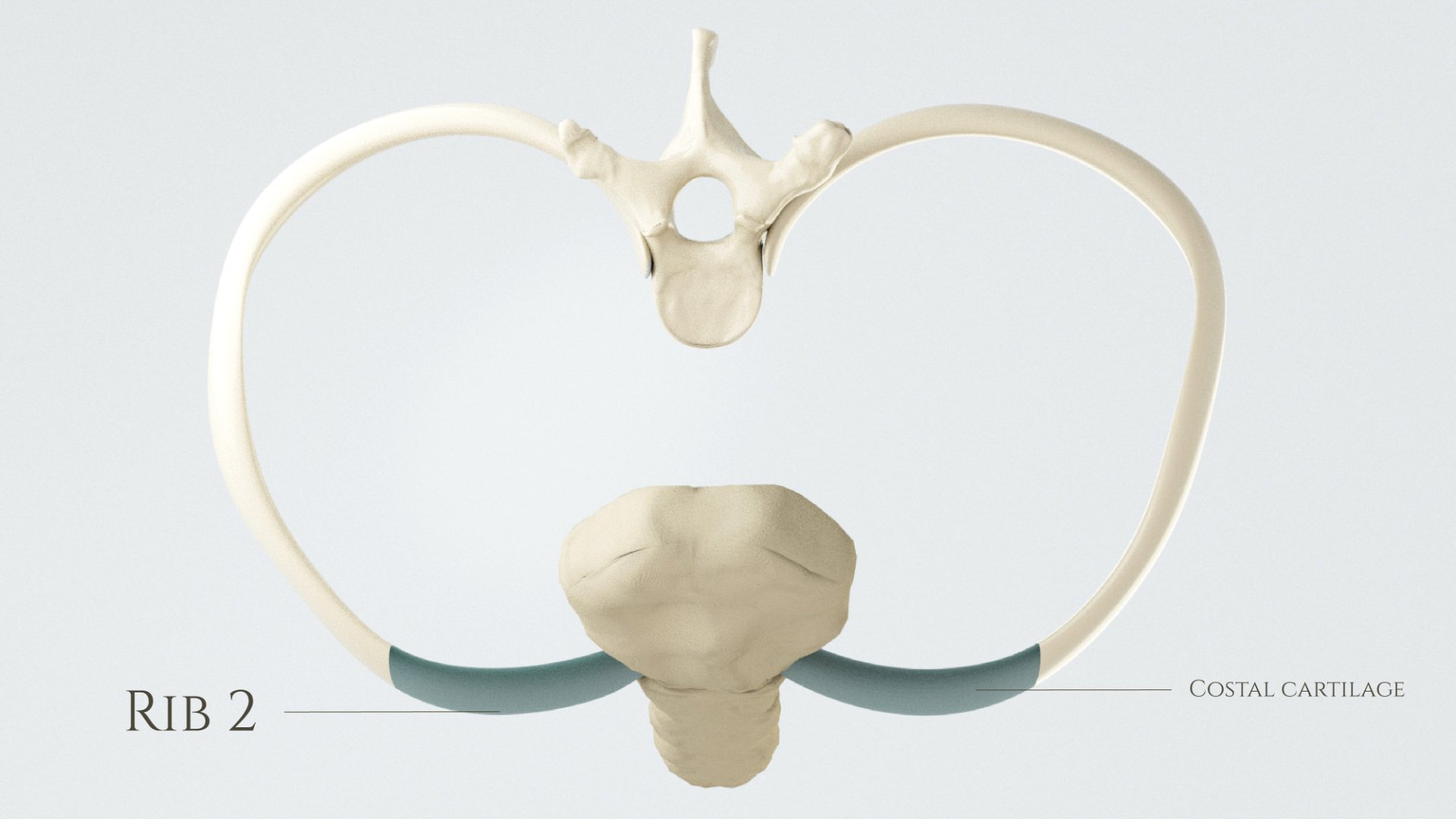
The Upper Skeleton
I was tasked to create a 3D anatomical model which includes the sternum, clavicle, scapula, humerus, ulna, radius, rib cage and the vertebral column, as ‘the upper skeleton’. Accurate anatomical model is very important when it comes to visualizing the anatomical relationship with other structures and showing the dimensions of the anatomy. Labels and annotations are used to describe the names of the structures.
Prior to modeling the skeleton, the dimensions, functionalities and the anatomical relationship are heavily researched to ensure the accuracy.
The dimensions (including body height anterior, height central, height posterior and intervertebral disk height) of all individual vertebrae are measured and followed the study from Iris Busscher's team: Busscher, I., Ploegmakers, J. J. W., Verkerke, G.J. et al. EurSpineJ 19, 1104-1114 (2010).
Anatomical modeling
Use of media: Autodesk Maya, Autodesk Mudbox, Adobe illustrator, Adobe Photoshop, 3D slicer, Adobe Substance Painter


Process
Here are some of the procedures I used in the 3D modeling process.
Sketching
Texturing and Individual look development, lighting
Overall look development, color grading

Elbow Joint
3D modeling | Graphic design | Anatomical visualization

This piece of work is focusing on the anterior and posterior views of the elbow joint. Bone textures and materials are added on the surface of 3 key structures of the arm: The humerus, radius and ulna. The joint is painted with an additional later to show the area of the cartilage.
The work acts as the thumbnail or introduction piece of an anatomical modeling project.
Use of media: Autodesk Maya, Mudbox, Adobe Illustrator, Photoshop
Descending Aorta
3D modeling | Graphic design | Anatomical visualization
This piece of work is focusing on the anterior and posterior views of the elbow joint. Bone textures and materials are added on the surface of 3 key structures of the arm: The humerus, radius and ulna. The joint is painted with an additional later to show the area of the cartilage.
The work acts as the thumbnail or introduction piece of an anatomical modeling project.
Use of media: Autodesk Maya, Mudbox, Adobe Illustrator, Photoshop
Acute Appendicitis
3D modeling | graphic design | molecular visualization

A vertical poster design showcasing the MoA of ibuprofen on pain management in patients with acute appendicitis. NSAIDs like ibuprofen, block the active site of cyclooxygenase (COX), the enzyme that converts arachidonic acid to prostaglandin. When the injured cells release more Arachidonic acid, COX enzyme thus converts them to prostaglandin (PGE2). Prostaglandin is a lipid compound in the cell membrane that mediate pain and inflammatory process.
A portrait illustration is added into the poster to mimic the real life situation. The poster is targeting to educated audience who have some biochemical knowledge, such as undergraduate students, and first-year medical students.
Use of media: Autodesk Maya, ChimeraX, PDB, Adobe Illustrator, Adobe Photoshop
Molecular visualization
“This piece of work is presented and displayed in the annual conference of the Association of Medical Illustrators (AMI). ”
Sketch and rendering

Color palette
Clothes
Background
Cell membrane
COX2
shadow
COX2
Photogrammetric Anteater

This piece of work demonstrated the techniques to use photogrammetry to make 3D models. Cinema 4D and Zbrush are used to clean up the models and clarify the textures of the Anteater.
Use of media: Autodesk Maya, Zbrush, Cinema 4D, Redshift
